Rembrandt’s Anatomical Portraits
By Siobhan M. Conaty, Ph.D.
There is a fascinating relationship between the 17th century Dutch master, Rembrandt van Rijn (1606-1669), and those in the fields of the health sciences and medical education. Rembrandt maintained both friendly and professional relationships with doctors throughout his career, and posthumously, health care professionals continue to remain intrigued and engaged with his works.[1] Multiple interpretations and analyses have been published in medical journals with topics that span from Rembrandt’s anatomy portraits, diagnoses of ocular issues and depression, to the depiction of aging and sickness in his self-portraits. [2] Anatomy serves as the foundation of the rehabilitation sciences. Closely examining two of Rembrandt’s anatomical paintings can provide students and practitioners with a new understanding of the foundation upon which rehabilitation sciences is based.
Rembrandt initially began his education as a philosophy student at the University of Leiden, but left in 1620 to study as an apprentice to the renowned artist Jacob van Swanenburgh. By 1631 he was established enough to set up his own studio in Amsterdam. He was only twenty-six years old when he was commissioned to paint the group portrait, The Anatomy Lesson of Dr. Nicolaes Tulp (1632) (Fig.1) by the Amsterdam Guild of Surgeons. Dr. Nicolaes Tulp was a prominent physician and respected civic leader who had recently become the director of anatomy at the Guild of Surgeons. At this time, the Church officially condemned dissection, but allowances were made for the sake of science — the guild had permission to conduct one dissection a year on a condemned criminal. The criminal in this case was Adriaan Adriaans (alias Aris Kindt), who was convicted and hanged for “grave assault” while attempting to steal a man’s cloak.[3] Rembrandt depicted Dr. Tulp in action, dissecting the cadaver while lecturing to seven other guild surgeons in attendance. As was the custom of the time, the surgeons paid to be included in this commission and so for the sake of history and commemoration, the names of the participants are stated clearly on a paper held by the surgeon standing to the left of Dr. Tulp.
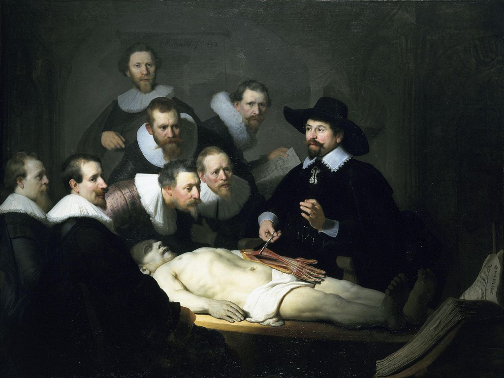
Rembrandt, The Anatomy Lesson of Dr. Nicolaes Tulp
Image credit: Mauritshuis, The Hague
Using forceps, Dr. Tulp conducts a lesson on the muscles and tendons of the hand and forearm. There is much discussion over a possible anatomical error committed by Rembrandt here, as the flexor digitorum superficialis (FDS) is shown laterally rather than medially.[4] Whether or not there is an error, the choice of “the hand” as a subject matter is significant. Dr. John Rupert Martin has noted that it was no coincidence that Dr. Tulp probes the muscle flexors that control the movement of the fingers while at the same time flexing his own left hand to demonstrate this same motion. While Rembrandt (and Dr. Tulp) demonstrate the literal use of the flexors, there is another layer of meaning occurring here. In the 16th century, famed Flemish anatomist Andreas Vesalius deemed it essential that an instructor should perform the dissection with his own hand while lecturing — rather than having someone else do the cutting. This was a new idea; one that Vesalius emphasized on the frontispiece of his landmark treatise De humani corporis fabrica of 1543 (The Fabric of the Human Body) (Fig.2). This woodcut illustrates a portrait of Vesalius dissecting the flexor superficialis of a forearm. It is very likely that the large book propped open at the foot of the cadaver in the Dr. Tulp painting is Vesalius’ Fabrica, thereby suggesting the importance of the theoretical and intellectual as well as the practical and clinical work of an anatomist. Rembrandt’s positioning of Dr. Tulp at this specific point of the dissection appears to honor Vesalius and his value of the work of the “hand” as a sign of important advancements in medical education in this Guild of Surgeons portrait. [5]
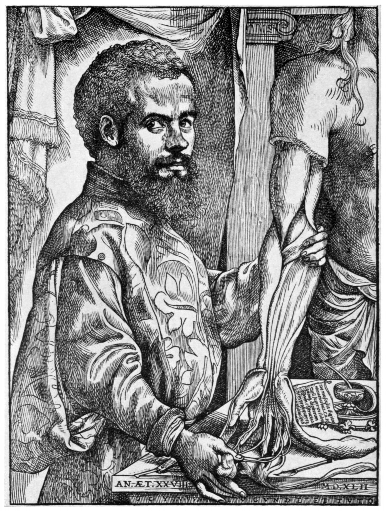
Frontispiece of Vesalius’ De humani corporis fabrica
Image credit: Art Resource
Scholars have noted that Rembrandt did not depict the scene as it occurred that day in 1632, although it is believed that he did attend the actual event.[6] Traditional dissections would begin with the perishable inner organs of the stomach and the brain, saving the extremities for the end. This was simply a practicality of the times, but by choosing to depict the dissection of the hand, Rembrandt was indeed making a specific choice (likely at Dr. Tulp’s request) and a clear reference to Vesalius. These yearly anatomical demonstrations were also open to the public, “physicians, surgeons, city magistrates, persons of note, even ladies, were invited.”[7] The events occurred in an anatomical theater, with the audience seated separately in a circular space around the dissection table. Rembrandt chose not to depict this larger scene; instead, breaking with 17th century Dutch tradition he depicted the surgeons in a more intimate grouping. There is a compelling psychological component to this small group as they look intently upon the cadaver, the medical textbook, and their instructor. Rembrandt extends the psychological invitation to the audience by having some of the figures make eye contact with us, the viewer. [8]
Rembrandt’s success after painting The Anatomy Lesson of Dr. Nicolaes Tulp, his first major commission, was swift. He became a celebrity of sorts; he was flooded with portrait requests (forty total in the two years after completing Dr. Tulp, compared to just three commissions the prior year) and introduced to important new patrons.[9] Tulp’s successor as head lecturer of anatomy at the Guild of Surgeons was Dr. Jan Deijman. The guild commissioned a second anatomy portrait in his honor in 1656. Painted nearly a quarter of a century later, Rembrandt’s The Anatomy Lesson of Dr. Deijman, (Fig.3) presents quite a contrast in composition.
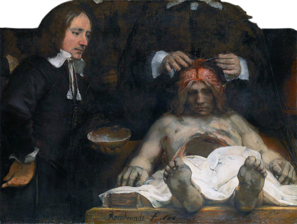
Rembrandt, The Anatomy Lesson of Dr. Deijman
Image credit: Amsterdam Museum, Amsterdam
Although the painting was partially destroyed in an 18th century fire, our understanding of the original composition is preserved in a surviving sketch of the completed painting. Dr. Deijman (whose head is no longer visible) stands as the central figure behind the cadaver. The cadaver is presented in stark foreshortening — a clear nod to the Italian Renaissance artist Andrea Mantegna’s Dead Christ (1480). Rembrandt, who never visited Italy, would have been familiar with Mantegna’s Dead Christ (1480) through printed reproductions. It is interesting to note that Mantegna’s Christ was criticized for looking too cold and heartless, akin to a corpse on a mortuary slab.
In The Anatomy Lesson of Dr. Deijman, Rembrandt placed the body of Joris Fonteijn (alias Black Jack), a recently executed criminal, on an operating table that seems to protrude out into viewer’s space – a visual trick to include the viewer in the scene. Rembrandt used foreshortening to psychologically invite the viewer in, we stand as part of the surgeon’s inner circle at the foot of the cadaver. This is in contrast to Dr. Tulp, where Rembrandt used eye contact to invite the viewer in – but only as observers or guests placed in the outer realm of the anatomical theater. Another variance in the Deijman painting is that Rembrandt reverts back to the traditional order of dissection of the time, the abdominal cavity lays open and empty while Dr. Deijman begins to work on the brain. An assistant holds the skullcap as Deijman moves the membranes to either side of the head in order to begin his probe of the brain. [10]
Both anatomy paintings, which were exhibited together in the Surgeon’s Guild until the 18th century fire, signal the cultural principles of the triumph of science and justice over crime – the fate of the executed criminals is made gruesomely clear.[11] Knowing these works were displayed along side each other; Rembrandt’s focus on the hands in the first portrait and the brain in the second offers a thought-provoking relationship between the two anatomy lessons. John Rupert Martin points out their complementary nature, one concerned with the hand, the “instrument of execution,” and the other concerned with the brain, the “center of thought and conception.”[12] When placed together, Rembrandt’s anatomical portraits seem to advocate for the mastery of both theory and practice in the study of the human body – two critical methods for success in the rehabilitation sciences.
References
[1] John Rupert Martin, Portraits of Doctors by Rembrandt and Rubens, Proceedings of the American Philosophical Society, Vol. 130, No. 1 (Mar., 1986), p.7.
[2] A small sample of articles on these topics:
Wiser I, Parnass AJ, Rachmiel R, Westreich M, Friedman T., Rembrandt’s Ocular Pathologies. Ophthal Plast Reconstr Surg. 2015 Jun 26.
Mondero NE1, Crotty RJ, West RW., Was Rembrandt strabismic? Optom Vis Sci. 2013 Sep;90(9):970-9.
Rosler R, Young P, The anatomy Lesson of Dr. Nicolaes Tulp: The beginning of a medical utopia. Rev Med Chil. 2011 Apr;139(4):535-41.
Friedman T, Westreich M, Lurie DJ, Golik A., Rembrandt–aging and sickness: a combined look by plastic surgeons, an art researcher and an internal medicine specialist. Isr Med Assoc J. 2007 Feb;9(2):67-71.
Marcus EL, Clarfield AM. Rembrandt’s late self-portraits: psychological and medical aspects. Int J Aging Hum Dev. 2002;55(1):25-49.
[3] Dolores Mitchell, Rembrandt’s “The Anatomy Lesson of Dr. Tulp”: A Sinner among the Righteous, Artibus et Historiae, Vol. 15, No. 30 (1994), pp. 145-152.
[4] See Robert Goldwyn’s discussion of the error in Nicolaes Tulp, Med Hist 1961; July; 5(3): 270-276, and LM Hove, S Young, JC Schrama, Dr. Nicolaes Tulp’s Anatomy Lecture, The Journal of the Norwegian Medical Association 2008; 128:716 – 9, for an opposing view.
[5] See John Rupert Martin’s Portraits of Doctors by Rembrandt and Rubens, Proceedings of the American Philosophical Society, Vol. 130, No. 1 (Mar., 1986), p.7 for further discussion of the Tulp/Vesalius connection.
[6] See Dolores Mitchell, p.151 and LM Hove, S Young, JC Schrama, 719.
[7] Robert Goldwyn, 271.
[8] See Aloïs Riegl and Benjamin Binstock, The Dutch Group Portrait, October, Vol. 74 (Autumn, 1995), pp. 3-35, for an in-depth discussion of Rembrandt’s composition.
[9] Robert Goldwyn, 272.
[10] For a recent analysis of Dr. Deijman’s anatomy lesson from a medical perspective, see Ijpma FF, Middelkoop NE, van Gulik TM. Rembrandt’s anatomy lesson of Dr. Deijman of 1656 dissected, Neurosurgery. 2013 Sep;73(3):381-5.
[11] David R. Smith, Inversion, Revolution, and the Carnivalesque in Rembrandt’s “Civilis” RES: Anthropology and Aesthetics, No. 27 (Spring, 1995), p.105.



 Member since 2019 | JM14274


NO COMMENT